Call us now
07315161963
Call us now
07315161963
HO 01 Composite Illustration of A Cell And Its Cytoplasmic organelles
HO 02 Different Types of Epithelia In Selected organs
HO 03 Composite Illustration of Loose Connective Tissue with Its Predominant Cells and Fibers
HO 04 Endochondral ossification, Illustrating the Progressive Stages of Bone Formation(from Cartilage Model
to Bone) and Including the Histology of A Section of Formed Bone
HO 05 Differentiation of A Pluripotential Hemopoietic Stem Into the Myeloid Stem Cell Line and Lymphoid
Stem Cell Line During Hemopoiesis
HO 06 Microscopic Illustrations of the Three Types of Muscles: Skeletal, Cardiac, and Smooth
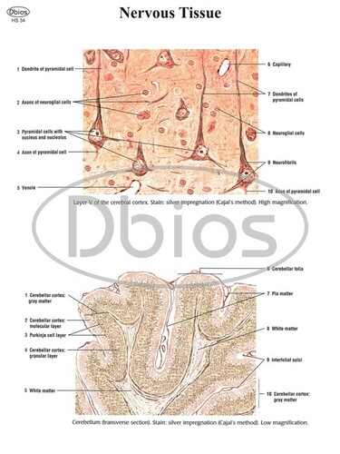
HO 07 The central nervous system is composed of the brain and spinal cord. A section of the brain and spinal
cord is illustrated here with their protective connective tissue layers called meninges (dura mater,
arachoid, and pia mater)
HO 08 The peripheral nervous system is composed of the cranial and spinal nerves. A cross - section of the
spinal cord is illustrated here with the characteristic features of the motor neuron and a cross-section
of a peripheral nerve. Also illustrated are types of neurons located in different ganglia and
organs outside of the central nervous system
HO 09 Comparison (transverse sections) of a muscular artery, large vein, and the three types of capillaries
HO 10 Location and distribution of the lymphoid organs and lymphatic channels in the body. Internal contents
of the lymph node and spleen are illustrated in greater detail
HO 11 Comparison between thin skin in the arm and thick skin in the palm, including contents of the
connective tissue dermis
HO 12 Salivary glands and their connections to the oral cavity, morphology of the tongue in cross - section,
and added detail of a taste bud
HO 13 Different types of acini (serous acini, mucous acini, and serous demilunes), different duct types
(intercalated,striated, and interlobular), and myoepithelial cells of a salivary gland
HO 14 Detailed illustration comparing the structural differences of the four layers ( mucosa, submucosa,
muscularis externa, and adventitia/serosa) in the wall of the esophagus and stomach
HO 15 Structural differences between the wall of the small intestine and large intestine, with emphasis on
different layers of the wall.
HO 16 A section from the liver and the pancreas is illustrated, with emphasis on the liver lobule and the duct
system of the exocrine pancreas.
HO 17 A section of the lung illustrated in three dimensions and in transverse section, with a emphasis on the
internal structure of the respiratory bronchiole and alveolar cells
HO 18 A sagittal section of the kidney showing the cortex and medulla, with blood vessels and the excretory
ducts, including the pelvis and the ureter and a histologic comparison of the blood vessels, the
different tubules of the nephron, and the collection ducts

Price:


|